YOUR GATEWAY TO CONFIDENTIAL REPRODUCTIVE & SEXUAL HEALTH ADVICE
Breast & Cervical Cancer

What is Breast Cancer?
Cancer begins when the body’s cell division functions abnormally. Normal cells grow and divide, forming new cells to take the place of dying cells. Sometimes, however, cells are created when they are not needed or the old cells do not die when they are supposed to.
The over-abundance of cells soon group together to create a mass or tumour. This tumour can be either benign (non-cancerous) or malignant (cancerous). A malignant tumour destroys the surrounding cells and tissue.
Cancers are most often named after the tumour growth’s initial location, and so breast cancer gets its name from problems occurring in breast cell division.
Most breast cancers begin in either the ducts (ductal carcinoma) or in the lobules (lobular carcinoma), although cancers can start in other breast tissues.
Types of Breast Cancer
The breasts are made of fat, glands, and connective (fibrous) tissue. The breast has several lobes, which are divided into lobules that end in the milk glands. Tiny ducts run from the many tiny glands, connect together, and end in the nipple.
Breast ducts are where 80% of breast cancers occur. This condition is called ductal cancer.
Cancer developing in the lobules is termed lobular cancer. About 10-15% of breast cancers are of this type.
Other less common types of breast cancer include inflammatory breast cancer, medullary cancer, mixed tumours, and a type of cancer involving the nipple termed Paget’s disease
Non-invasive Breast Cancer
Non-invasive breast cancer can be defined as cancer that does not spread from the breast lobules or ducts to the surrounding tissue or to other parts of the body.
Invasive breast cancer
Invasive breast cancer is cancer that has spread beyond the breast lobules of ducts to surrounding tissue.
When cancers spread into the surrounding tissues, they are termed infiltrating cancers. Cancers spreading from the ducts into adjacent spaces are termed infiltrating ductal carcinomas. Cancers spreading from the lobules are infiltrating lobular carcinomas.
Metastatic breast cancer
The most serious cancers are metastatic cancers. Metastasis means that the cancer has spread from the place where it started into other tissues distant from the original tumour site.
The most common place for breast cancer to spread is into the lymph nodes under the arm or above the collarbone on the same side as the cancer. Other common sites of breast cancer metastasis are the brain, the bones, and the liver.
Recurrent breast cancer
Recurrent breast cancer is cancer that comes back after treatment. It may recur in the breast or chest wall, or in any other part of the body.
What are the risk factors associated with developing Breast Cancer?
Age
- Age is the most significant risk factor
- Breast cancer is rare in women younger than 25 years and incidence increases with age
Genetics
- Family history is a risk factor, both maternal and paternal relatives are important. The lifetime risk is up to 4 times higher if a mother and sister are affected. The family history characteristics that suggest increased risk of cancer are as follows:
- Two or more relatives with breast or ovarian cancer
- Breast cancer occurring in an affected relative younger than 50 years
- Relatives with both breast cancer and ovarian cancer
- One or more relative with 2 cancers. Male relatives with breast cancer
- Breast Cancer Genes: About 5-10% of breast cancers are believed to be hereditary, as a result of mutations, or changes, in certain genes that are passed along in families.
Hormonal Causes
- Factors increasing the number of menstrual cycles increase the risk of breast cancer most probably due to long exposure to internal hormones such as estrogen. These factors includecharacteristics that suggest increased risk of cancer, and are as follows:
- nulliparity (never been pregnant)
- first full pregnancy when older than 30 years
- menarche when younger than 13 years
- menopause when older than 50 years
- not breastfeeding
- Hormone replacement therapy increases risk (1.35 times for 5 or more years use, normalizing 5 years from discontinuing).
- The use of oral contraceptive pills except progesterone only pills increases risk (1.24 times for 10 years use, normalizing 10 years from discontinuing).
Dietary and Lifestyle Causes
- Obesity, increased alcohol intake and a sedentary lifestyle, increase risk of breast cancer
Exogenous factors
- Irradiation, example excessive exposure to X-rays, particularly in the first decade of life, is associated with an increased risk of breast cancer.
- Certain metabolite of insecticides and pesticides increase risk of developing breast cancer.
Do I have Breast Cancer?
Early breast cancer has no symptoms. It is usually not painful.
Most breast cancer is discovered before symptoms are present, either by finding an abnormality on mammography (see below) or feeling a breast lump. A lump in the armpit or above the collarbone that does not go away, may be a sign of cancer.
Other possible symptoms are one sided, bloody nipple discharge, newly developed nipple inversion, or skin changes of the overlying breast such as redness and dimpling.
Most breast lumps are not cancerous. All breast lumps, however, need to be evaluated by a doctor.
Breast cancer develops over months or years. Once it is identified, however, a certain sense of urgency is felt about the treatment, because breast cancer is much more difficult to treat as it spreads. You should see your physician if you experience any of the following:
- Finding a breast lump
- Finding a lump in your armpit or above your collarbone that does not go away in two weeks or so
- Developing nipple discharge
- Noticing new nipple inversion or skin changes over the breast
- If an abnormality is found on your mammogram (see below), you should see your health-care provider right away to make a plan for further evaluation.
What tests do I need to determine if I have cancer? What is a mammogram?
Diagnosis of breast cancer usually is comprised of several steps, including examination of the breast, mammography, possibly ultrasonography or MRI, and, finally, biopsy. Biopsy is the only definitive way to diagnose breast cancer.
1) Examination of the Breast
A complete breast examination includes visual inspection and careful palpation (feeling) of the breasts, the armpits, and the areas around your collarbone.
During that exam, your physician may palpate a lump or just feel a thickening.
2) Mammography
Mammograms are x-rays of the breast that may help define the nature of a lump. Mammograms are also recommended for screening to find early cancer. Usually, it is possible to tell from the mammogram whether a lump in the breast is breast cancer, but no test is 100% reliable. Mammograms are thought to miss as many as 10-15% of breast cancers.
A mammogram is done in conjunction with other tests since a mammogram alone is often not enough to evaluate a lump.
3) Ultrasound
Ultrasound of the breast is often done to evaluate a breast lump in younger patients. Ultrasound waves create a “picture” of the inside of the breast. It can demonstrate whether a mass is filled with fluids (cystic) or solids. Cancers are usually solid, while many cysts (fluid filled lump) are benign. Ultrasound might also be used to guide a biopsy or the removal of fluid.
4) MRI
MRI may provide additional information and may clarify findings which have been seen on mammography or ultrasound. MRI is not routine for screening of cancer but may be recommended in special situations.
5) Biopsy
The only way to diagnose breast cancer with certainty is to biopsy the tissue in question. Biopsy means to take a very small piece of tissue from the body for examination and testing by a pathologist. Pathologists are physicians who are specially trained in diagnosing diseases by looking at cells and tissues under a microscope to determine if cancer is present.
A number of biopsy techniques are available.
Fine-needle aspiration consists of placing a needle into the breast and sucking out some cells to be examined by a pathologist. This technique is used most commonly when a fluid-filled mass is identified and cancer is not likely.
Core-needle biopsy is performed with a special needle that takes a small piece of tissue for examination. Usually the needle is directed into the suspicious area with ultrasound or mammogram guidance. This technique is being used more and more because it is less invasive than surgical biopsy. It obtains only a sample of tissue rather than removing an entire lump. Occasionally, if the mass is easily felt, cells may be removed with a needle without additional guidance.
Surgical biopsy is done by making an incision in the breast and removing the piece of tissue. Certain techniques allow removal of the entire lump.Regardless of how the biopsy is taken, the tissue will be reviewed by a pathologist.
If a cancer is diagnosed on biopsy, the tissue will be tested for hormone receptors.
Receptors are sites on the surface of tumour cells that bind to estrogen or progesterone. In general, the more receptors, the more sensitive the tumour will be to hormone therapy. There are also other tests (for example, measurement of HER-2/neu receptors) that may be performed to help characterize a tumour and determine the type of treatment that will be most effective for a given tumour.
What is the treatment of breast cancer?
Awareness of breast cancer symptoms and signs of breast cancer may help for early diagnosis. Breast cancer treatment is dependent on stage and type of breast cancer. Treatment may include surgery, radiation, hormonal agents, or chemotherapy and is usually divided into medical or surgical treatment.
Medical Treatment
Many women have treatment in addition to surgery, which may include radiation therapy, chemotherapy, or hormonal therapy. The decision about which additional treatments are needed is based upon the stage and type of cancer, the presence of hormonal and/or HER-2/neu receptors, and patient health and preferences.
Radiation therapy is used to kill tumour cells if there are any left after surgery. Radiation is a local treatment and therefore works only on tumour cells that are directly in its beam. It is usually given five days a week over five to six weeks with each treatment taking only a few minutes. Radiation therapy is painless and has relatively few side effects. However, it can irritate the skin or cause a mild burn.
Chemotherapy consists of the administration of medications that kill cancer cells or stop them from growing.
Chemotherapy is usually given in “cycles.” Each cycle includes a period of intensive treatment lasting a few days or weeks followed by a week or two of recovery. Most people with breast cancer receive at least two, more often four, cycles of chemotherapy to begin with. Tests are then repeated to see what effect the therapy has had on the cancer.
Chemotherapy differs from radiation in that it treats the entire body and thus may target stray tumour cells that may have migrated from the breast area.
The side effects of chemotherapy are well known. Side effects depend on which drugs are used. Many of chemotherapy drugs have side effects that include loss of hair, nausea and vomiting, loss of appetite, fatigue, and low blood cell counts. Low blood counts may cause patients to be more susceptible to infections, to feel sick and tired, or to bleed more easily than usual. Medications are available to treat or prevent many of these side effects.
Hormonal therapy may be given because breast cancers (especially those that have high amounts of estrogen or progesterone receptors) are frequently sensitive to changes in hormones. Hormonal therapy may be given to prevent recurrence of a tumour or for treatment of existing disease.
Surgery
Surgery is generally the first step after the diagnosis of breast cancer. The type of surgery is dependent upon the size and type of tumour and the patient’s health and preferences.
Lumpectomy Lumpectomy involves removal of the cancerous tissue and a surrounding area of normal tissue. This is not considered curative and is done in association with other medical therapy such as radiation therapy with or without chemotherapy or hormonal therapy.
Simple mastectomy removes the entire breast but no other structures. If the cancer is invasive, this surgery alone will not cure it. It is a common treatment for DCIS, a noninvasive type of breast cancer.
Modified radical mastectomy removes the breast and the axillary (underarm) lymph nodes but does not remove the underlying muscle of the chest wall. Although additional chemotherapy or hormonal therapy is almost always offered, surgery alone is considered adequate to control the disease if it has not metastasized.
Radical mastectomy involves removal of the breast and the underlying chest wall muscles, as well as the underarm contents. This surgery is no longer done because current therapies are less disfiguring and have fewer complications.
People who have been diagnosed with breast cancer need careful follow-up care for life. Initial follow-up care after completion of treatment is usually every three to six months for the first two to three years.
Follow-up includes careful breast examination, mammography, blood work, and, possibly, a chest x-ray or other studies such as bone scans or CT scans.
Where do I go to get treatment?
Breast Cancer is treated at all major tertiary care hospitals in Pakistan. Breast Cancer treatment is done through a multidisciplinary approach that involves for the most part surgery and oncology departments. In tertiary care hospitals, Breast clinics provide specialized care to breast cancer patients.
Breast clinics are conducted by the General Surgery department which deals with surgical aspects of breast cancer. Oncology department of tertiary care hospitals or specialized nuclear and radiation therapy units are responsible for the medical treatment of breast cancer such as radiotherapy, hormonal and chemotherapy.
Can I prevent Breast Cancer?
The most important risk factors for the development of breast cancer are sex, age, and genetics. Because women can do nothing about these risks, regular screening is recommended in order to allow early detection and thus prevent death from breast cancer.
Regular screening includes breast self-examination, clinical breast examination, and mammography.
For women who are menstruating, the best time for examination is immediately after the monthly period.
For women who are not menstruating or whose periods are extremely irregular, picking a certain date each month seems to work best.
Instruction in the technique of breast self-examination can be obtained from a physician associated with a breast clinic.
Clinical breast examination: The frequency of the clinical breast examination depends on whether you are at high risk or at a low risk of developing breast cancer. The American Cancer Society recommends a breast examination by a trained health-care provider once every three years starting at age 20 years, and then yearly after age 40 years.
Mammograms are recommended every one to two years starting at age 40 years. For women at high risk for the development of breast cancer, mammogram screening may start earlier, generally 10 years prior to the age at which the youngest close relative developed breast cancer.
Lifestyle modification is encouraged. Physically active women may have a lower risk of developing breast cancer. All women are encouraged to maintain normal body weight, especially after menopause and to limit excess alcohol intake. Hormone replacement should be limited in duration if it is medically required.
Cervical Cancer:
Cervical cancer develops in a woman’s cervix (the entrance to the uterus from the vagina).
Almost all cervical cancer cases (99%) are linked to infection with high-risk human papillomaviruses (HPV), an extremely common virus transmitted through sexual contact.
Although most infections with HPV resolve spontaneously and cause no symptoms, persistent infection can cause cervical cancer in women.
Cervical cancer is the fourth most common cancer in women. In 2018, an estimated 570 000 women were diagnosed with cervical cancer worldwide and about 311 000 women died from the disease.
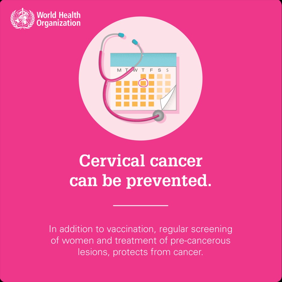
Effective primary (HPV vaccination) and secondary prevention approaches (screening for and treating precancerous lesions) will prevent most cervical cancer cases.
When diagnosed, cervical cancer is one of the most successfully treatable forms of cancer, as long as it is detected early and managed effectively. Cancers diagnosed in late stages can also be controlled with appropriate treatment and palliative care.
With a comprehensive approach to prevent, screen and treat, cervical cancer can be eliminated as a public health problem within a generation.

